Cellular Division
One of the characteristics of living things is the ability to replicate and pass on genetic information to the next generation. Cell division in individual bacteria and archaea usually occurs by binary fission. Mitochondria and chloroplasts also replicate by binary fission, which is evidence of the evolutionary relationship between these organelles and prokaryotes.
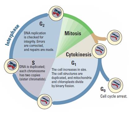
Cell division in eukaryotes is more complex. It requires the cell to manage a complicated process of duplicating the nucleus, other organelles, and multiple chromosomes. This process, called the cell cycle, is divided into three parts: interphase, mitosis, and cytokinesis (Figure 1). Interphase is separated into three functionally distinct stages. In the first growth phase (G1), the cell grows and prepares to duplicate its DNA. In synthesis (S), the chromosomes are replicated; this stage is between G1and the second growth phase (G2). In G2, the cell prepares to divide. In mitosis, the duplicated chromosomes are separated into two nuclei. In most cases, mitosis is followed by cytokinesis, when the cytoplasm divides and organelles separate into daughter cells. This type of cell division is asexual and important for growth, renewal, and repair of multicellular organisms.
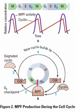
Cell division is tightly controlled by complexes made of several specific proteins. These complexes contain enzymes called cyclin-dependent kinases (CDKs), which turn on or off the various processes that take place in cell division. CDK partners with a family of proteins called cyclins. One such complex is mitosis-promoting factor (MPF), sometimes called maturation-promoting factor, which contains cyclin A or B and cyclin-dependent kinase (CDK). (See Figure 2a.) CDK is activated when it is bound to cyclin, interacting with various other proteins that, in this case, allow the cell to proceed from G2 into mitosis. The levels of cyclin change during the cell cycle (Figure 2b). In most cases, cytokinesis follows mitosis.
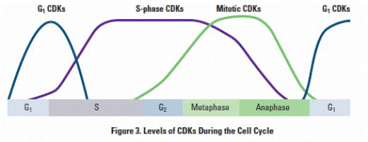
As shown in Figure 3, different CDKs are produced during the phases. The cyclins determine which processes in cell division are turned on or off and in what order by CDK. As each cyclin is turned on or off, CDK causes the cell to move through the stages in the cell cycle.
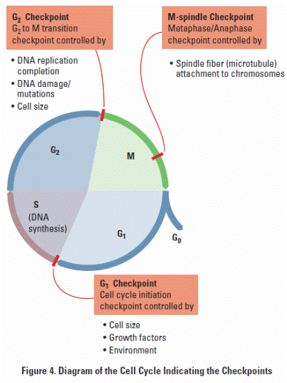
 Cyclins and CDKs do not allow the cell to progress through its cycle automatically. There are three checkpoints a cell must pass through: the G1 checkpoint, G2 checkpoint, and the M-spindle checkpoint (Figure 4). At each of the checkpoints, the cell checks that it has completed all of the tasks needed and is ready to proceed to the next step in its cycle. Cells pass the G1 checkpoint when they are stimulated by appropriate external growth factors; for example, platelet-derived growth factor (PDGF) stimulates cells near a wound to divide so that they can repair the injury. The G2 checkpoint checks for damage after DNA is replicated, and if there is damage, it prevents the cell from going into mitosis. The M-spindle (metaphase) checkpoint assures that the mitotic spindles or microtubules are properly attached to the kinetochores (anchor sites on the chromosomes). If the spindles are not anchored properly, the cell does not continue on through mitosis. The cell cycle is regulated very precisely. Mutations in cell cycle genes that interfere with proper cell cycle control are found very often in cancer cells.
Prelab Questions
1. How did you develop from a single-celled zygote to an organism with trillions of cells? How many mitotic cell divisions would it take for one zygote to grow into an organism with 100 trillion cells?
2. How is cell division important to a single-celled organism?
3. What must happen to ensure successful cell division?
4. How does the genetic information in one of your body cells compare to that found in other body cells?
5. What are some advantages of asexual reproduction in plants?
6. Why is it important for DNA to be replicated prior to cell division?
7. How do chromosomes move inside a cell during cell division?
8. How is the cell cycle controlled? What would happen if the control were defective?
9. What is the role of Cyclin and CDK?
Counting Cells and Analyzing Data
1. Observe the cells at high magnification (400–500 X).
2. Look for well-stained, distinct cells.
3. Within the field of view, count the cells in each phase. Repeat the counts in two other root tips.
4. Collect the class data for each group, and calculate the mean and standard deviation for each group.
5. Compare the number of cells from each group in interphase and in mitosis.
Group Data
Tip |
Number of Cells |
||
Interphase |
Mitotic |
Total |
|
1 |
|
|
|
2 |
|
|
|
3 |
|
|
|
Total |
|
|
|
Mean |
|
|
|
Standard Deviation |
|
|
|
![]()
Number of Cells |
Group |
Group |
Group |
Group |
Group |
Group |
Group |
Group |
Average |
Standard Deviation |
I |
|
|
|
|
|
|
|
|
|
|
M |
|
|
|
|
|
|
|
|
|
|
Effects of Environment on Mitosis
Scientists reported that a fungal pathogen, may negatively affect the growth of soybeans (Glycine max). Soybean growth decreased during three years of high rainfall, and the soybean roots were poorly developed. Close relatives of R. anaerobis are plant pathogens and grow in the soil. A lectin-like protein was found in the soil around the soybean roots. This protein may have been secreted by the fungus. Lectins induce mitosis in some root apical meristem tissues. In many instances, rapid cell divisions weaken plant tissues.
You have been asked to investigate whether the fungal pathogen lectin affects the
number of cells undergoing mitosis in a different plant, using root tips.
1. What is your experimental hypothesis? Your null hypothesis? Are these the same?
2. How would you design an experiment with onion bulbs to test whether lectins
increase the number of cells in mitosis?
3. What would you measure, and how would you measure it?
4. What would be an appropriate control for your experiment and experimental variables that must be controlled?
Upon completion of the experiment, the following data was collected.
2. Use the data from Table 2 above to calculate the totals using the formulas found in Table 2. (For example, A equals the number of interphase cells in the control group.)
3. Use the totals from Table 2 to calculate the expected values (e) using the formulas
from Table 3.
4. Enter the observed values (o) from Table 2 and expected values (e) from Table 3 for each group into Table 4. Calculate the chi-square (χ2) value for the data by adding together the numbers in the right column.
5. Compare this value to the critical value in Table 5.
1. The degrees of freedom (df) equals the number of treatment groups minus one
multiplied by the number of phase groups minus one. In this case, there are two
treatment groups (control, treated) and two phase groups (interphase, mitosis);
therefore df = (2 - 1) (2 - 1) = 1.
2. The ρ value is 0.05, and the critical value is 3.84. If the calculated chi-square value is greater than or equal to this critical value, then the null hypothesis is rejected. If the
calculated chi-square value is less than this critical value, the null hypothesis is not
rejected.
Loss of Cell Cycle Control in Cancer
The last question is the focus of this part of the lab. With your group, form a hypothesis as to how the chromosomes of a cancer cell might appear in comparison to a normal cell and how those differences are related to the behavior of the cancer cell.
For each of the following cases, research and look at pictures of the chromosomes (karyotype) from normal human cells. Compare them to pictures of the chromosomes from HeLa cancer cells. For each case, count the number of chromosomes in each type of cell, and discuss their appearance. Then answer the following questions.
Case 1: HeLa cells
HeLa cells are cervical cancer cells isolated from a woman named Henrietta Lacks.
Her cells have been cultured since 1951 and used in numerous scientific experiments.
Henrietta Lacks died from her cancer not long after her cells were isolated. Lacks’s
cancer cells contain remnants of human papillomavirus (HPV), which we now know
increases the risk of cervical cancer.
Case 2: Philadelphia Chromosomes
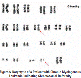
In normal cells, mitosis usually is blocked if there is DNA damage. Sometimes, though, DNA damage makes cells divide more often. Certain forms of leukemia have a unique feature called a Philadelphia chromosome. Look at the karyotype of leukemia cells in Figure 5, and answer the following questions:
Source: http://www.wuhsd.org/site/handlers/filedownload.ashx?moduleinstanceid=12709&dataid=30167&FileName=Cellular%20Division.doc
Web site to visit: http://www.wuhsd.org
Author of the text: not indicated on the source document of the above text
If you are the author of the text above and you not agree to share your knowledge for teaching, research, scholarship (for fair use as indicated in the United States copyrigh low) please send us an e-mail and we will remove your text quickly. Fair use is a limitation and exception to the exclusive right granted by copyright law to the author of a creative work. In United States copyright law, fair use is a doctrine that permits limited use of copyrighted material without acquiring permission from the rights holders. Examples of fair use include commentary, search engines, criticism, news reporting, research, teaching, library archiving and scholarship. It provides for the legal, unlicensed citation or incorporation of copyrighted material in another author's work under a four-factor balancing test. (source: http://en.wikipedia.org/wiki/Fair_use)
The information of medicine and health contained in the site are of a general nature and purpose which is purely informative and for this reason may not replace in any case, the council of a doctor or a qualified entity legally to the profession.
The following texts are the property of their respective authors and we thank them for giving us the opportunity to share for free to students, teachers and users of the Web their texts will used only for illustrative educational and scientific purposes only.
All the information in our site are given for nonprofit educational purposes
The information of medicine and health contained in the site are of a general nature and purpose which is purely informative and for this reason may not replace in any case, the council of a doctor or a qualified entity legally to the profession.
www.riassuntini.com